Lumbar facet arthrosis
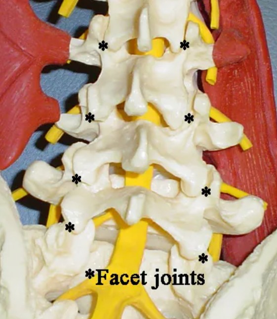
The facet joints are on the posterior (back side) aspect of the spine, and are to the right and the left from the midline. These joints, which have a sliding motion to allow the spine to flex forward and lean backward, have surfaces covered by cartilage, like on the end of a chicken bone. These joints are indicated in the picture on the right by the asterisk (*) on the photograph of the spine model.
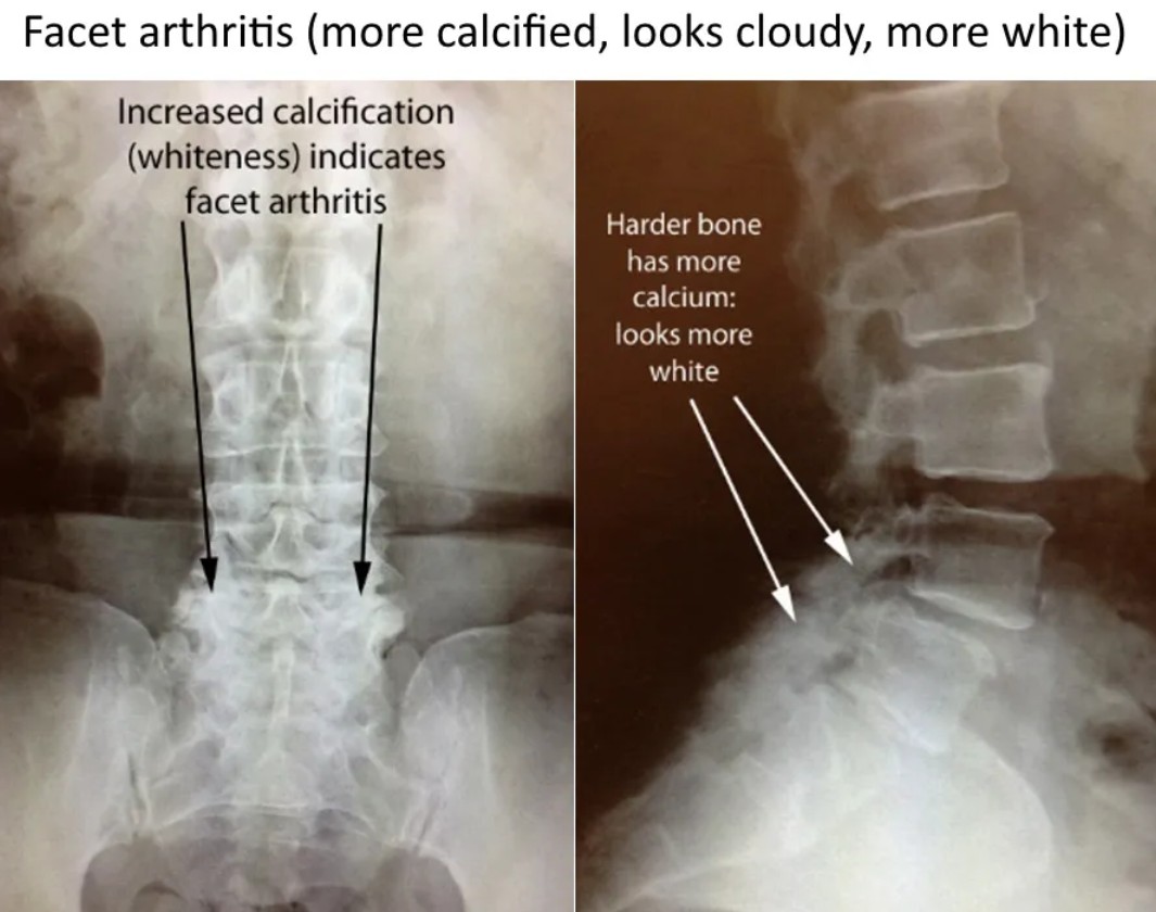
When the cartilage wears away, usually in a process of degeneration (wear and tear), the surfaces become rough with bone rubbing on bone. The body tries to fix this problem…by making more bone. Therefore, arthritic joints appear more calcified, more white, on Xrays.
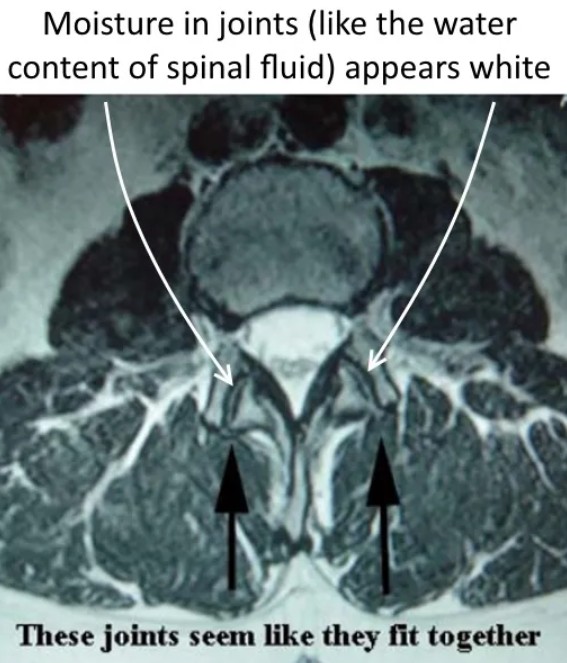
Cross sectional imaging
A better way to look at these joints is with a three-dimensional study, like an MRI or CT scan, that can make images in different planes/directions. In this cross sectional view which shows normal facet joints, notice that it appears that the two bones that form the facet joint fit nicely together. There is a thin line of normal appearing joint fluid between the surfaces of the facet joints. This thin line is present since the cartilage in the joint has a higher degree of moisture/water content than the underlying bone.
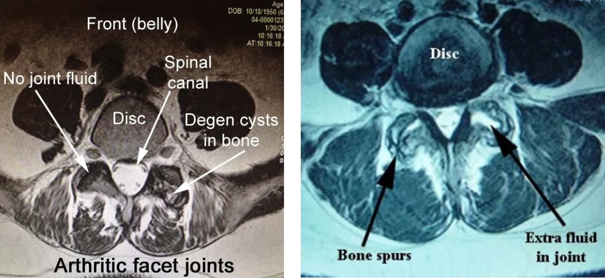
Below are examples of abnormal facet joints shown in MRI scans. In the image below left, the lack of fluid in the facet along with the degenerative cysts can be seen.
In the image below right, due to degenerative changes and large bone spurs forming, there is no longer normal sliding motion in these joints. On one side, since the two components of the joint no longer fit together, there is too much fluid in that joint space, which is not a normal finding. It is not hard to imagine that the patient would have pain with spinal flexion and extension.
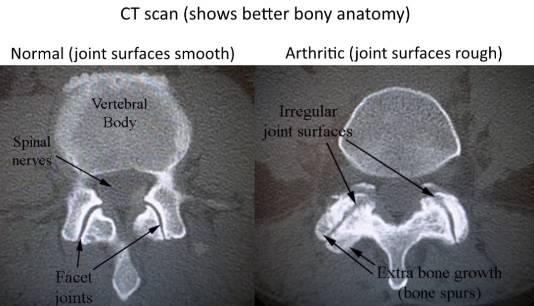
Facet arthritis is illustrated even better with a CT scan. On the side with arthritis, it's almost like the joints, which should be smooth, have rusted.
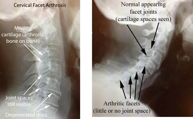
Cervical facet arthrosis
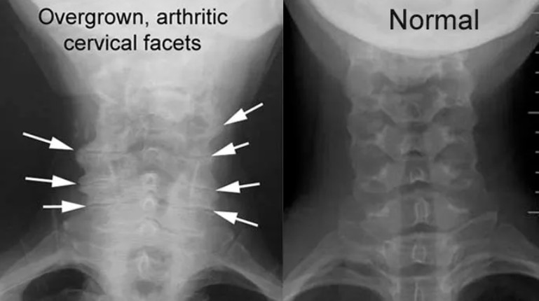
The facet joints in the back part of the cervical spine can also wear out in a similar manner.
In the images shown here, which are AP views (looking from front to back), arthritic and enlarged facet joints can be compared to more normal appearing joints.
Treatment options
In most cases, the pain specialists will treat facet related pain, first with diagnostic blocks which help to confirm from where the pain is being generated. If they do the blocks and the patient has no relief, we lack convincing evidence that further intervention would help.
However, if the patient has relief with the blocks, they could be an candidate for a procedure to make a small burn near the nerves to these joints, a procedure called radiofrequency ablation (RFA). Often, a successful RFA can give relief for 3-12 months.